Thursday, December 25, 2014

Respiratory Examination


Introduction :-

In medicine, the respiratory examination is performed as part of a physical examination, or when a patient presents with a respiratory problem (dyspnea (shortness of breath), coughchest pain) or a history that suggests a pathology of the lungs. It is very rarely performed in its entirety or in isolation, most commonly it's merged with the cardiac examination.


The respiratory exam has conventionally been split into different stages:
  • Position of the patient and the environment.
  • Inspection of the patient and respiratory effort.
  • Palpation of the patient's anterior and posterior chest.
  • Percussion of the patient's anterior and posterior chest walls.
  • Auscultation of the patient's anterior and posterior chest walls.
One method to remember the steps of the examination is through the mnemonic PIPPA.
When accompanying other physicians or students, medical staff typically report as they examine a patient. Examples of a normal examination may include:
  • adjustment of environment
  • "on inspection, effort is normal"
  • "no tenderness to palpation", "fremitus WNL"
  • "clear to percussion bilaterally" or "CTP B"
  • "clear to auscultation bilaterally" or "CTA B"; "no R/R/W" or "no rhonchirales or wheezes"

Positioning and Environment :-
  • In the respiratory examination, the patient is asked to sit upright on an examination table, with arms at the side. Adequate lighting is ensured, and the patient is asked to expose the chest. Later in the examination, when the back is examined, the patient is usually asked to move the arms forward so that the scapulae are not in the way of examining the upper lung fields. These fields are intended to correlate with the lung lobes, and are thus tested on the anterior and posterior chest walls (the front and back of the patient's thorax).
Inspection:-

The examiner then observes the patient's respiratory rate, which is typically conducted under the pretext of some other exam, so that patient does not subconsciously increase their baseline respiratory rate. Signs of respiratory distress may include:
  • Cyanosis, a bluish tinge of the extremities (peripheral cyanosis), or of tongue (central cyanosis).
  • Pursed-lip breathing
  • Accessory muscle use,including the scalene and intercostal muscles.
  • Diaphragmatic breathing, paradoxical movement of the diaphragm outwards during inspiration.
  • Intercostal indrawing.
  • Decreased chest-chest movement on the affected side.
  • An increased Jugular venous pressure, indicating possible right heart failure
Chest wall abnormalities are also examined, and may include:
  • Kyphosis, abnormal anterior-posterior curvature of the spine
  • Scoliosis, abnormal lateral curvature of the spine
  • Barrel chest, - chest wall increased anterior-posterior; normal in children; typical of hyperinflation seen in COPD
  • Pectus excavatum - sternum sunken into the chest
  • Pectus carinatum - sternum protruding from the chest
As well as the patient's respiratory rate, the pattern of breathing is also noted:
  • An acidotic patient will have more rapid breathing to compensate, known as Kussmaul breathing.
  • Cheyne–Stokes respiration, which is alternating breathing in high frequency and low frequency from brain stem injury. It can be seen in newborn babies which is sometimes physiological (normal). Also observe for retractions seen in asthmatics. Retractions can be supra-sternal, where the accessory muscles of respirations of the neck are contracting to aid inspiration. Retractions can also be intercostal, in which there is visible contraction of the intercostal muscles (between the ribs) to aid in respiration. These are signs of respiratory distress.
The physician then typically inspects the fingers for cyanosis and clubbing.
Tracheal deviation is also examined.

Palpation:-

The physician then places both palms on the posterior lung fields, asking the patient to count 1 to 10. The physician aims to feel for vibrations and compare the right/left lung fields. If the patient has a consolidation, (for example caused by pneumonia), the vibration will be louder at that part of the lung. This is because sound travels faster through denser material than air.
If a patient has pneumonia, palpation may reveal increased vibration and dullness on percussion. If there is pleural effusion, palpation should reveal decreased vibration and there will be 'stony dullness' on percussion.
The examiner then tests for
  • Tracheal deviation, whether trachea is in centred or not, indicating enlargement or collapse of a lung field.
  • Tactile fremitus, with the patient asked to say boy-O-boy or ninety-nine, and the physician using the ulnar aspect of their hand to feel changes in sound conduction.
  • Respiratory expansion, indicating whether lung expansion is equal.
  • the position of the apex beat to check if there has been deviation of the heart.

Percussion:-

The physician attempts to examine changes in density of the lung fields by examining its resonance.
Specifically, percussion is performed with the middle finger striking the middle phalanx of the other middle finger of the other hand. The sides of the chest are compared. This is performed symmetrically on all lung fields, on the anterior and posterior chest walls.
Examples of alterations in density may include pleural effusion and pneumothorax. The sound is described as tympanic if there is a pneumothorax because air will stretch the pleural membranes. Conversely, if there is fluid between the pleural membranes, the percussion will be dampened and sound muffled.

Auscultation:-

 The physician then auscultates the respiratory sounds over the lung fields, listening to the fields through a stethoscope. This is conducted while the patient is breathing, noting normal breath sounds and any abnormalities including:
  • Wheezes, describing a continuous musical sound on expiration or inspiration. A wheeze is the result of narrowed airways. Common causes include asthma and emphysema
  • Rhonchi (an increasingly obsolete term) characterised by low pitched, musical bubbly sounds heard on inspiration and expiration. Rhonchi are the result of viscous fluid in the airways.
  • Crackles or rales. Intermittent, non-musical and brief sounds heard during inspiration only. They may be described as fine(soft, high-pitched) or coarse (louder, low-pitched). These are the result of alveoli opening due to increased air pressure during inspiration. Common causes include congestive heart failure.
  • Stridor a high-pitched musical breath sound resulting from turbulent air flow in the larynx or lower in the bronchial tree. It is not to be confused with stertor. Causes are typically obstructive, including foreign bodies, croupepiglottitis, tumours, infection and anaphylaxis.
  • Appropriate ratio of inspiration to expiration time (expiration time increased in COPD)
  • Bronchial or vesicular breath sounds.
Lastly an assessment of transmitted voice sounds is performed.

Differential Diagnosis Of Respiratory Diseases :-


Take Care Of Your Body,

It's The Only Place You Have To Live In.

With Best Regards, Karnav Thakkar :) :)

Thursday, December 18, 2014

Abdominal Examination

Introduction :-
The abdominal exam, in medicine, is performed as part of a physical examination, or when a patient presents with abdominal pain or a history that suggests an abdominal pathology.
The abdominal exam has conventionally been split into different stages:
  • Positioning of the patient and their environment.
  • Inspection of the patient and their anterior and posterior abdomen.
  • Auscultation of the abdomen with a stethoscope.
  • Palpation of the patient's abdomen and its organs.
  • Percussion of the patient's abdomen.
  • Special tests inspecting for signs of various liver diseases.
When accompanying other physicians or students, medical staff typically report as they examine a patient. An example normal examination may include:
  • adjustment of environment
  • "on inspection, the abdomen is non-distended and there are no signs of liver disease"
  • "bowel sounds are present"
  • "the abdomen is soft and non-tender, with no organomegaly"
  • "no rebound tenderness"
Positioning and Environment :-
  • Position is patient should be supine and the bed or examination table should be flat. The patient's hands should remain at his/her sides with his/her head resting on a pillow. If the neck is flexed, the abdominal musculature becomes tensed and the examination made more difficult. Allowing the patient to bend his/her knees so that the soles of their feet rest on the table will also relax the abdomen.
  • Lighting is adjusted so that it is ideal.
  • Draping is patient should be exposed from the pubic symphysis below to the costal margin above - in women to just below the breasts. Some surgeons would describe an abdominal examination being from nipples to knees.
  • Although physicians have had concern that giving patients pain medications during acute abdominal pain may hinder diagnosis and treatment.

Inspection:-

The abdominal exam typically begins with the examiner inspecting the patient, paying particular attention to any signs of liver disease. These signs (stigmata) may include
The patient is also examined for:

Auscultation:-

  • Auscultation refers to the use of a stethoscope by the examiner to listen to the abdomen.
  • Auscultation is performed prior to palpation of the abdomen as it is least likely to elicit pain.
  • The doctor warms the diaphragm of the stethoscope, and listens to the bowel sounds. Some controversy exists as to the length of time required to confirm or exclude bowel sounds, with suggested durations up to five minutes. Bowel obstruction may present with grumbling bowel sounds or high-pitched noises. Absence of sounds may be caused by peritonitis.
  • The examiner also typically listens to the two renal arteries for bruits by listening in each upper quadrant, adjacent to and above the umbilicus. Bruits heard in the epigastrium that are confined to systole are considered normal.

Percussion:-


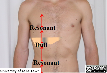
Percussion is performed by knocking the middle finger against the phalanx of the middle finger of the opposing hand, which rests against the surface of the abdomen in each of the nine areas tested. Percussion can elicit a painful response in the patient, and may also reveal whether there is abnormal levels of fluid in the abdomen. Organomegaly may also be noted, including gross splenomegaly (enlargement of the spleen), hepatomegaly (enlargement of the liver), and urinary retention.
The examiner, when percussing for organomegaly, percusses in a particular manner:
  • percuss the liver from the right iliac region to right hypochondrium
  • percuss for the spleen from the right iliac region to the right hypochondrium and the left iliac to the left hypochondrium.
Examination of the spleen

Palpation:-

The examiner typically palpates all nine areas of the patient's abdomen. This is typically performed twice, lightly and then deeply.
On light palpation, the examiner tests for any palpable mass, rigidity, or pain.
On deep palpation, the examiner is testing for and organomegaly, including enlargement of the liver and spleen.
Reactions that may indicate pathology include:
  • guarding, describing muscle contraction as pressure is applied.
  • rigidity, indicating peritoneal inflammation.
  • rebound, pain on release
  • hernial orifices if positive cough impulses.

Other & Special Maneuvers:-


  • Examination of pelvic lymph nodes
  • Digital rectal exam - Abdominal examination is not complete without a digital rectal exam.
  • Pelvic examination only if clinically indicated.
Special manevures may also be performed, to elicit signs of specific diseases. These include


Take Care Of Your Body,

It's The Only Place You Have To Live In.

With Best Regards, Karnav Thakkar :) :)

Thursday, December 11, 2014

General Examination

Introduction :-

  • The general examination of the patient must be done systematically, noting the following -
  1. Built
  2. Body proportions
  3. Nutrition
  4. Decubitus
  5. Skin, hair and nails
  6. Clubbing
  7. Cyanosis
  8. Jaundice
  9. Pallor
  10. Lymphadenopathy
  11. Edema
  12. Vertebral Column
  13. Thickened nerves
  14. Joints
  15. Temperature
  16. Pulse
  17. Blood Pressure

1.Built :- 
  • Built is the skeletal structure in relation to age and sex of the individual as compared to a normal person.


Tall Stature :-

  • A child is considered to be tall when the height is greater than 2 standard deviations above the mean for the age. Gigantism is the term applied when the patient's height is greatly in excess of the normal for his age before fusion of epiphysis. There is no fixed height to continue a giant, but in adults, it is applied for individuals with a height of more than 61/2 Ft.


Short Stature :-

  • Dwarfism is the term applied when the patient's height is 2 standard deviations less than that for his/her age and sex. mid-parental height usually determines the final height.


2.Body Proportions :- 

  • Normally, in adults, the height of the person is equal to the length of arm span. The upper segment ( from vertex to the pubic symphisis ) is equal to the lower segment ( from pubic symphisis to the heel ).
  • In infants, the upper segment is greater than the lower segment and the height is greater than the arm span. This infantile type of body proportion persists in achondroplasia, cretinism and juvenile myxedema.
  • The reverse of infantile body proportion i.e. arm span greater than height and lower segment greater than upper segment occurs in eunuchoidism, marfan's syndrome, homocystinuria, klinefelter's syndrome and frohlich's syndrome.

3.Nutrition :- 

  • A normal person is well nourished as regards proteins, fats, carbohydrates, vitamins and minerals. certain clinical signs help to diagnose deficiency of one or more of these nutrients.
  1. Proteins :- Hypoproteinemia causes rough skin and later edema of feet and brittle hair.
  2. Fats :- Fat malnutrition leads to cachexia with hollowing of cheeks, loss of the shape of hips ( due to loss of fats ), flat abdomen and absent fat over the subcutaneous tissues of the elbows.
  3. Carbohydrates :- Carbohydrate malnutrition is difficult to detect clinically because there is gluconeogenesis from fats or proteins.
  4. Vitamins :- These can be fat soluble ( vitamin A, D, E, K ) or water soluble (rest).
  5. Minerals :- Deficiency of two minerals can be diagnosed clinically. Iron deficiency causes koilonychia and pallor whereas calcium deficiency causes tetany.


4.Decubitus :- 

  • Decubitus or the posture a patient adopts when lying in bed often gives a valuable diagnostic clue.

  1. Hemiplegia :- The patient lies in bed with one side immobile, the affected arm flexed and the affected leg externally rotated and extended.
  2. Meningitis and tetanus :- The patient has neck stiffness and opisthotonos.
  3. Colic :- In renal, biliary or intestinal colic, the patient is markedly restless and tossing and turning in bed in agony.
  4. Acute inflammatory abdominal disease :- The patient lies on his back quietly with legs drawn up.
  5. Cardiorespiratory embarrasment :- The patient is more comfortable in sitting-up position. This position is also assumed in abdominal distention and ascites when intra-abdominal pressure is raised.
  6. Pneumonia and pleurisy :- The patient is most comfortable lying on the affected side because the movement on the affected side is restricted.


5.Skin,Hair & Nails :- 


Examination of skin often gives important clues to local or systemic diseases. The following features should be noted :

  • Color :- It may be pale, flushed, cyanosed, yellow etc.
  • Pigmentation :- Pigmentation may occur in several diseases. some common medical conditions associated with pigmentation are :
  • Endocrine : Addison's disease, cushing's disease, thyrotoxicosis. 
  • Deficiency : pellagra, kwashiorkor, megaloblastic anemia. 
  • Infections : kala azar, chronic malaria, secondary syphilis, tuberculosis, leprosy, HIV etc. 
  • Metabolic : Hemochromatosis. 
  • Skin disease : neurofibromatosis, lichen planus, acanthosis nigricans etc. 
  • Miscellaneous :  Malignancy, pernicious anemia, exposure to sunrays or radiations. 

  • Hypopigmentation :- Hypopigmented patches may occur in leprosy, leukoderma, albinism, fungal infections of skin, etc.


  • Eruptions : various types of eruptions may occur as follows:


Examination of Hair :


Examination of Nails :


6.Clubbing :- 


7.Cyanosis :- 


8.Jaundice :- 


9.Pallor :- 

  • pallor is paleness of skin and mucous membrane either as a result of diminished circulating red blood cells or diminished blood supply.

Paleness should be distinguished from other causes of prominent white skin:
  • Fair skin is genetically determined skin hue with low concentration of skin pigment (melanin) in the skin. This skin hue is common in people in north European countries (Germany, Great Britain, Ireland, Scandinavian countries).
  • Absence of skin tan from sun avoidance.
  • Myxedema (in hypothyroidism) – swelling of under-skin tissues causes pale appearance of the skin.
  • Albinism is a rare genetic disorder with partial or complete lack of melanin in the skin, hair and iris of the eye. Affected persons have white skin and hair, and red iris.
  • Vitiligo is a patchy loss of skin color due to destruction of pigment cells (melanocytes) from an unknown cause.

Everyday Causes of Paleness

Paleness does not always mean you are ill.
  • When exposed to low environmental temperature, your face, palms or other body parts may become pale because of narrowing (constriction) of the small skin arteries as part of a body’s heat-saving process.
  • When you keep your arms or legs above the level of the heart for a minute, they may become pale (and numb or tingling), since the power of the heart can not efficiently pump the blood into the limbs against the force of gravitation.
  • Skipped meal and resulting drop of glucose blood level, or dehydration, may trigger adrenalin release and constriction of your skin arteries.
  • In exertion or fear, blood is redirected from the skin to muscle arteries. Your skin may remain pale for several minutes after exertion.

Sudden Paleness

Health disorders, causing sudden paleness (within minutes to hours) all over the body:
  • Orthostatic hypotension – temporary fall of blood pressure after standing up after prolonged sitting or lying.
  • Stomach upset from wrong food combination, alcohol or food poisoning
  • Dehydration from insufficient drinking, excessive sweating, vomiting, diarrhea
  • Acute infection (usually with fever)
  • Fainting (vasovagal syncope) due to strong pain, emotions, heat or unpleasant sensations
  • Motion sickness
  • Allergy to drugs
  • Rapid stomach emptying (dumping syndrome)
  • Migraine
  • Heat stroke
  • Hypothermia
  • Heart failure due to heart attack, arrhythmia, infective endocarditis or other heart disorder, when the heart can not efficiently pump the blood into the circulation
  • Low blood sugar (hypoglycemia), common in insulin-dependent diabetics after an exercise, skipped meal or insulin overdose
  • Blood loss due to external or internal bleeding (in car accidents, shooting or stitch injuries), heavy menstrual bleeding, surgery
  • Hypothermia
  • Shock - a sudden, deep fall of blood pressure - due to poisoning, severe infection, burns, severe blood loss
  • Side effect of medications:
    • warfarin, corticosteroids, aspirin and other anti-rheumatic drugs may cause intestinal bleeding
    • iron poisoning
  • Drug overdose: amphetamine (speed), cocaine
  • Chemical poisoning (pesticides), plant poisoning (Atropa belladonna)
  • Death

Sudden Paleness in Limbs

Health disorders, causing sudden paleness in limbs:

Long Lasting Paleness

Paleness lasting from few weeks to several years and affecting the whole body may result from:
  • Low blood pressure, insufficient to keep small skin arteries open
  • Anemia due to iron or vitamin B12 and folate deficiency due to
    • low intakevegetarians, irregular diet, starvation, alcoholism
    • impaired absorptionCrohn’s or celiac disease
    • intestinal parasites
    • intestinal bleeding: colorectal cancer, ulcerative colitis
    • heavy menstrual bleeding
    • hemolysis
    • drugs phenytoin and methotrexate
    • chronic kidney disease
    • chemotherapy
    • cancer in advanced stage
    • chronic hepatitis or liver cirrhosis
    • pregnancy
  • Hypertension
  • Chronic heart failure after heart attack, heart valve disorders, etc.
  • Side effect of medications:
    • warfarin, aspirin, ibuprofen, naproxen, corticosteroids may cause intestinal bleeding
    • iron poisoning
  • Drug abuse: amphetamine (speed), cocaine
  • Thrombosis and other blood clotting disorders
  • Leukemia, lymphoma, Hodgkin disease
  • Hypopituitarism - impaired production of pituitary hormones, mainly due to pituitary adenoma

Long Lasting Paleness in Limbs

  • Chronic arterial occlusion
  • Carpal tunnel syndrome
  • Acromegaly

Paleness in Children

Disorders, commonly or mainly causing paleness in children:
  • Dehydration
  • Stress from travel
  • Childhood diseases with fever
  • Iron deficiency anemia
  • Malnutrition due to starvation or lack of proteins in the diet
  • Rheumatic fever
  • Congenital heart disorders
  • Cystic fibrosis
  • Genetic metabolic disorders: phenylketonuria
10.Lymphadenopathy :- 

  • Lymphadenopathy is inflammatory or non-inflammatory enlargement of lymphnodes.


11.Edema :- 


12.Vertebral column :- 


13.Thickened nerves :- 

  • It may be due to 
  • leprosy
  • Neurofibromatosis
     
  •  Diabetes 
  •  Amylodosis 
  •  Charcot marie tooth syndrome 
  •  Sarcoidosis 
  •  Refsum's Disease 
  •  Rusy Levy Syndrome 
  •  Dejerine sotta's syndrome 
  •  Idiopathic hypertrophic neuropathy 
  •  CIDP ( Chronic Inflammatory Demylinating polyneuropathy )
 14.Joints :- 

15.Temperature :- 


16.Pulse :- 


17.Blood Pressure :- 


Take Care Of Your Body,

It's The Only Place You Have To Live In.

With Best Regards, Karnav Thakkar :) :)